The medical archives holds over 1,000,000 photographic items dating from the late 19th century to the present. These collections reflect the daily use of photography within an academic health institution and encompass many genres, including biomedical, clinical, documentary, corporate, architectural, and portrait photography. The collections also encompass many historic and current photographic processes, including cased photographs, card photographs, photomicrographs, glass plate negatives and slides, albumen, platinum, and gelatin silver prints, color photographic prints, transparencies, and slides, digitally produced prints, and born-digital photographs.
The photograph collections are arranged either topically or by office or department of origin. To browse the finding aids to our major photograph collections, explore the list below. To search for specific types of photographs among all collections, use the advanced search tab on the catalog search page. On the first search row, select Format Type in the first column, Exact in the second column, and Image in the third column. Use the remaining rows to search using keywords, titles, or subjects. Please note that although many photographs are digitized and viewable through the online catalog, many are not. To search for only photographs that have been digitized, select the digital content only button after you search. To access photographs that are not digitized, you may schedule an appointment to view the photographs in our reading room, or request photocopies or scans of photographs for a fee. Visit our reproduction fees page for more information. Appointments and other reference inquiries can be made through our online registration form.
Major Photographic Collections
Explore finding aids to our major photographic collections. Additional photographic series may be found in larger personal paper collections and institutional records.
- Portraits of Individuals Photograph Collection
Includes over 2,300 photographic portrait files of faculty, staff, and other individuals associated with the health divisions of Johns Hopkins.
- Buildings Photograph Collection
Includes exterior, interior, and construction photographs of buildings across Johns Hopkins’ medical campuses.
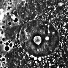
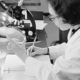
- Biomedical Photograph Collection
Includes photomicrographs and clinical photographs from various sources.
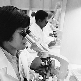
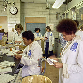
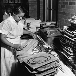
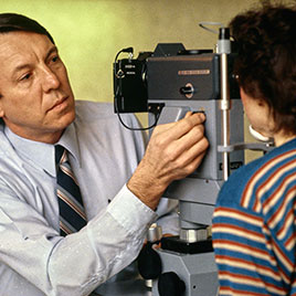
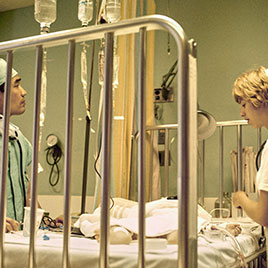
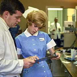
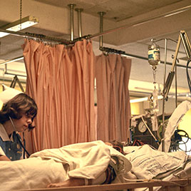
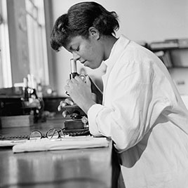
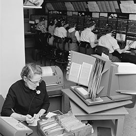
- People at Work Photograph Collection
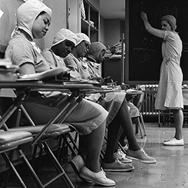
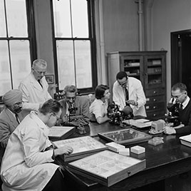
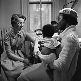
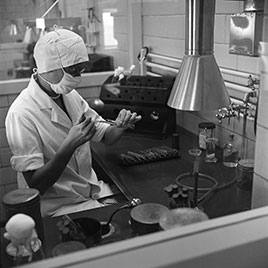
Includes active photographs of Johns Hopkins physicians, nurses, technicians and other employees at work. - Johns Hopkins University Homewood Photography Collection
Includes more recent photographs of events and portraits of Johns Hopkins Medicine, Nursing, and Public Health faculty, staff, and students, photographed for Johns Hopkins publications and websites.
- Office of Public Affairs Photograph Collection
Includes photographs taken for Johns Hopkins publications and promotional use, and entire negative files of Johns Hopkins staff photographers, dating predominantly from 1956-1996. - Office of Marketing and Communications Publications Photographs
Marketing and Communications is the successor to the Office of Public Affairs. Includes photographs taken for Johns Hopkins Medicine publications, dating predominantly from 1988-2007. - The Johns Hopkins Hospital and School of Medicine Photograph Collection
Includes annual departmental group portraits, School of Medicine class portraits, individual student record portraits, and photograph albums.
- Johns Hopkins Nursing Photograph Collection
Includes annual class group portraits and individual student record portraits for the Johns Hopkins Hospital School of Nursing, and photograph albums.
